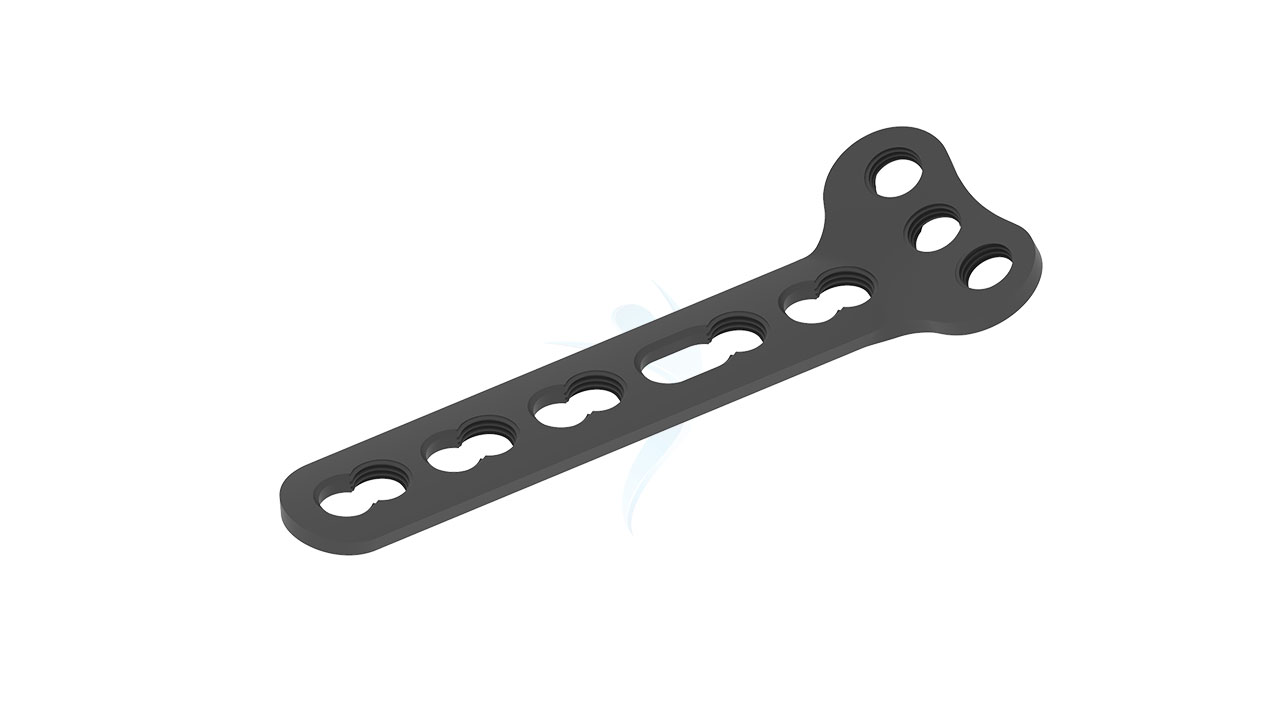
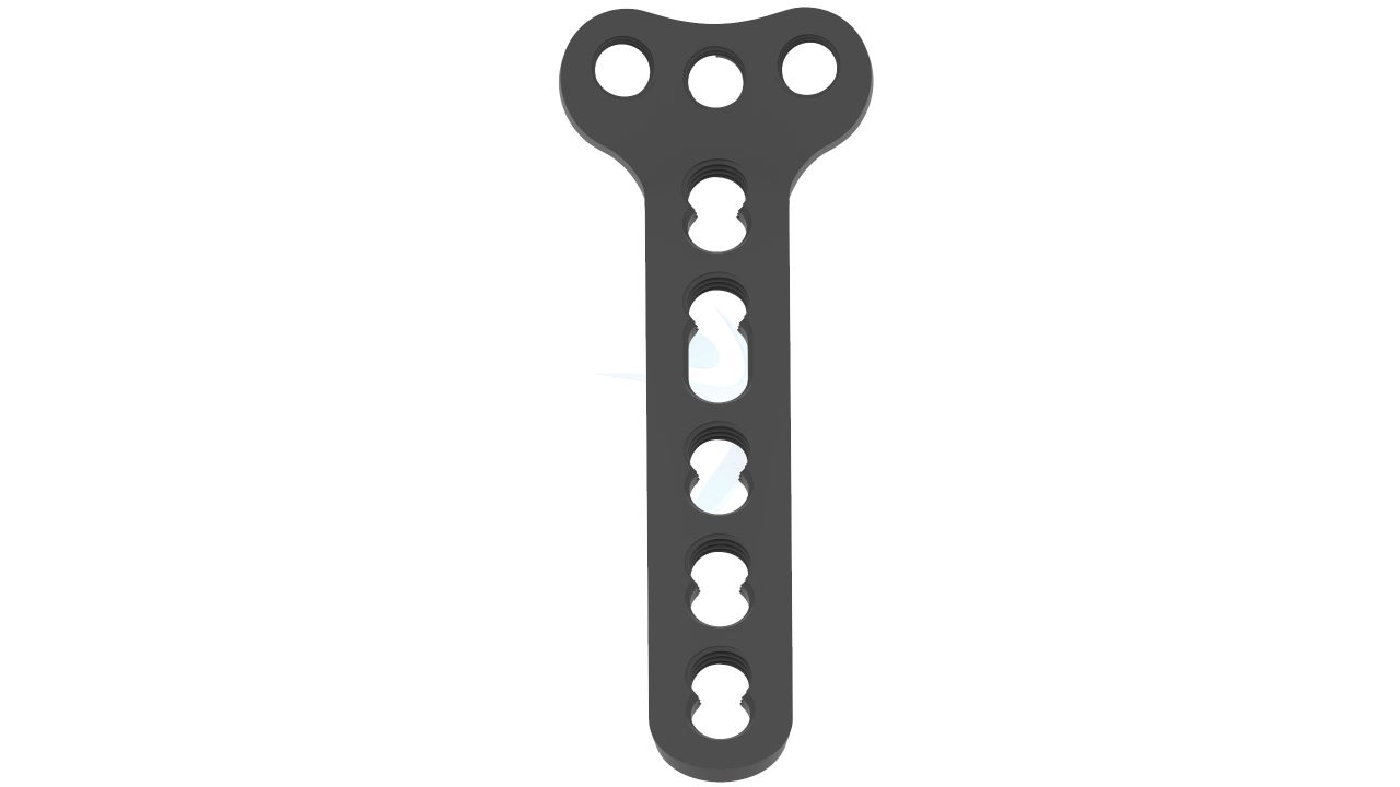
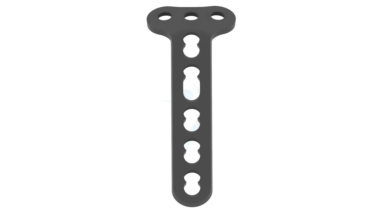
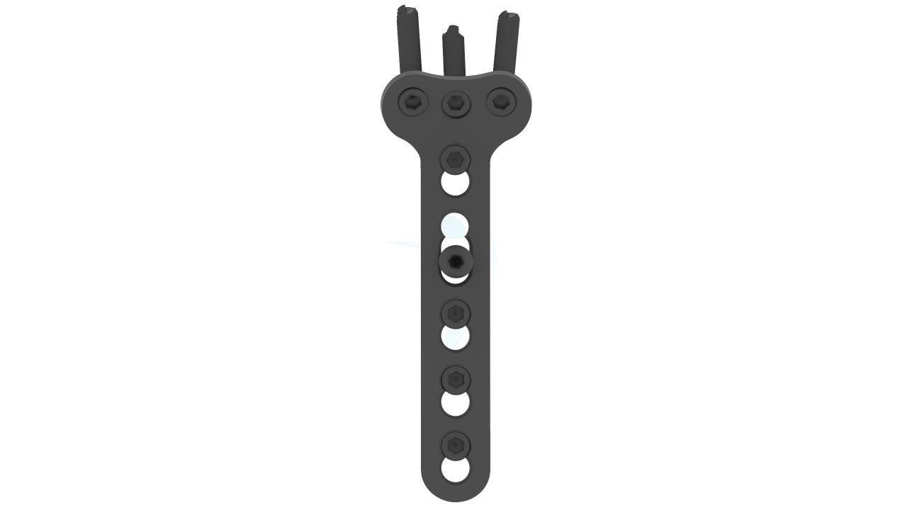
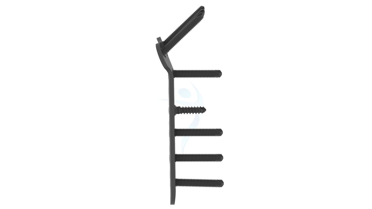
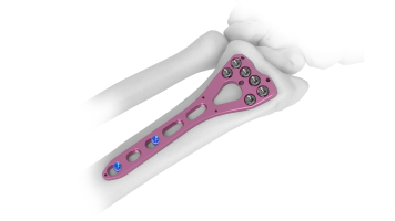
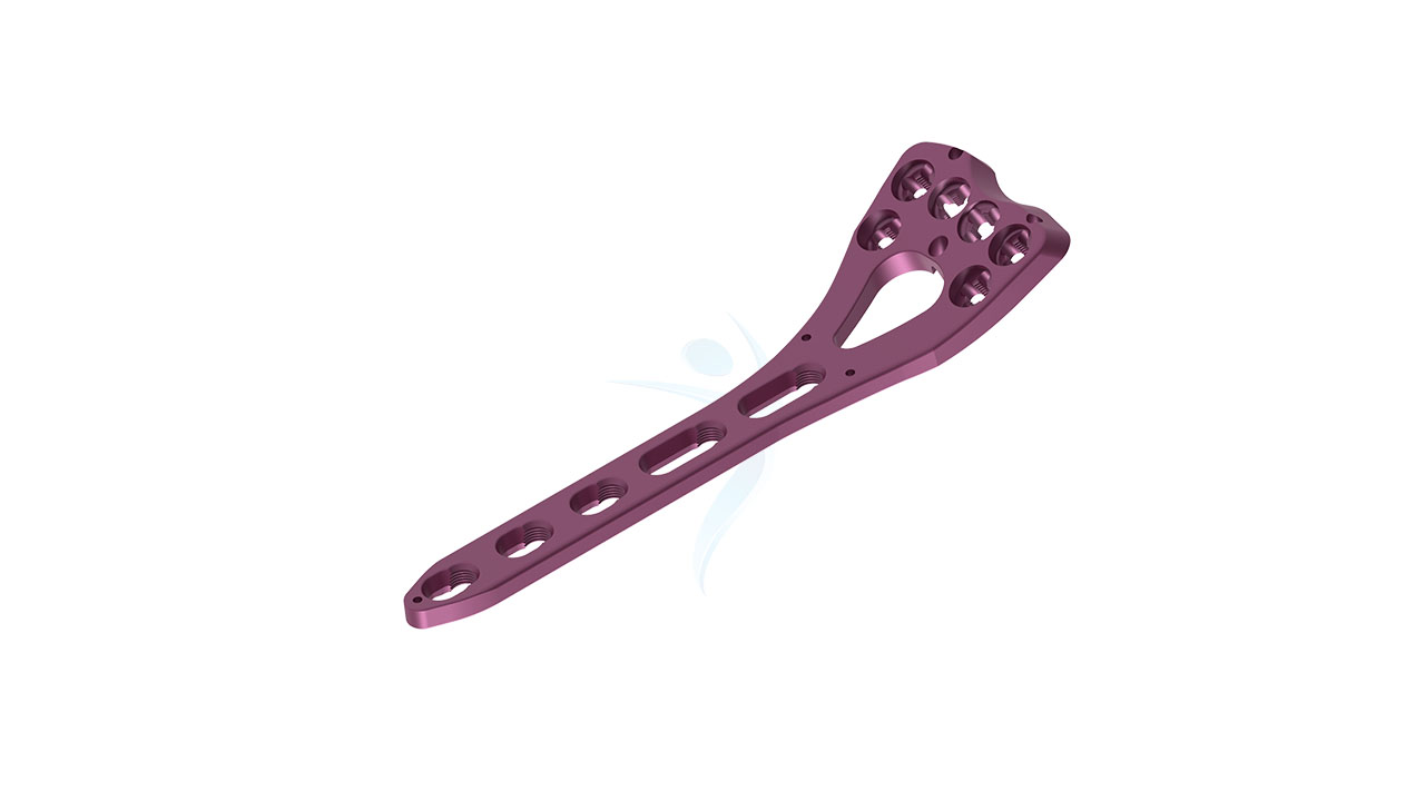
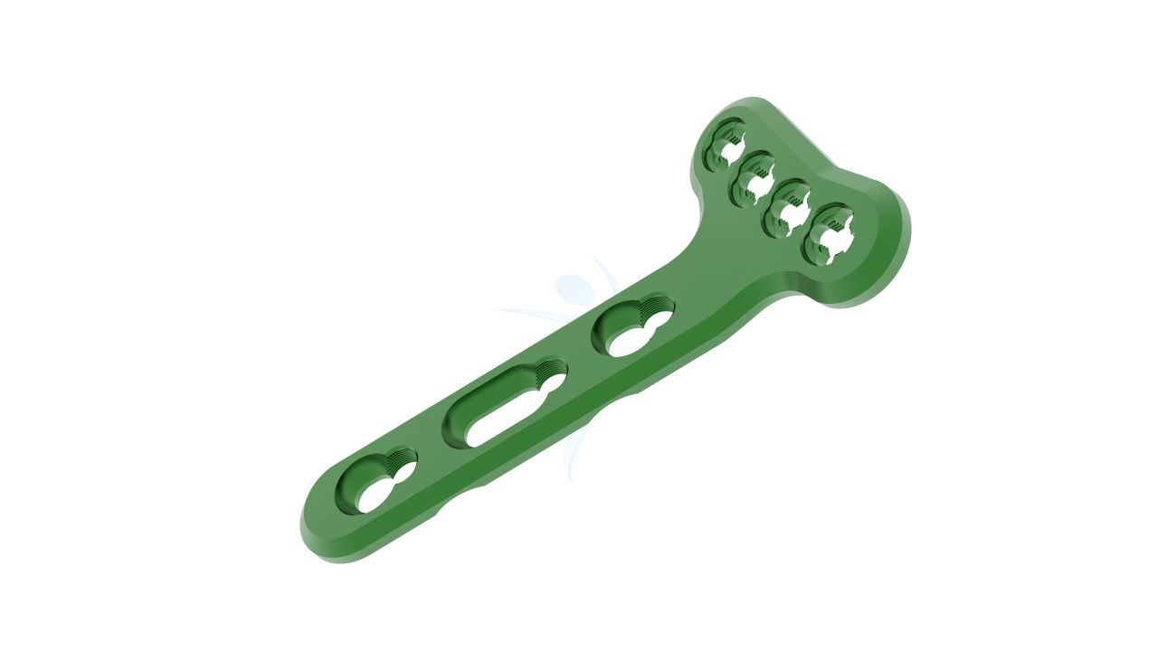
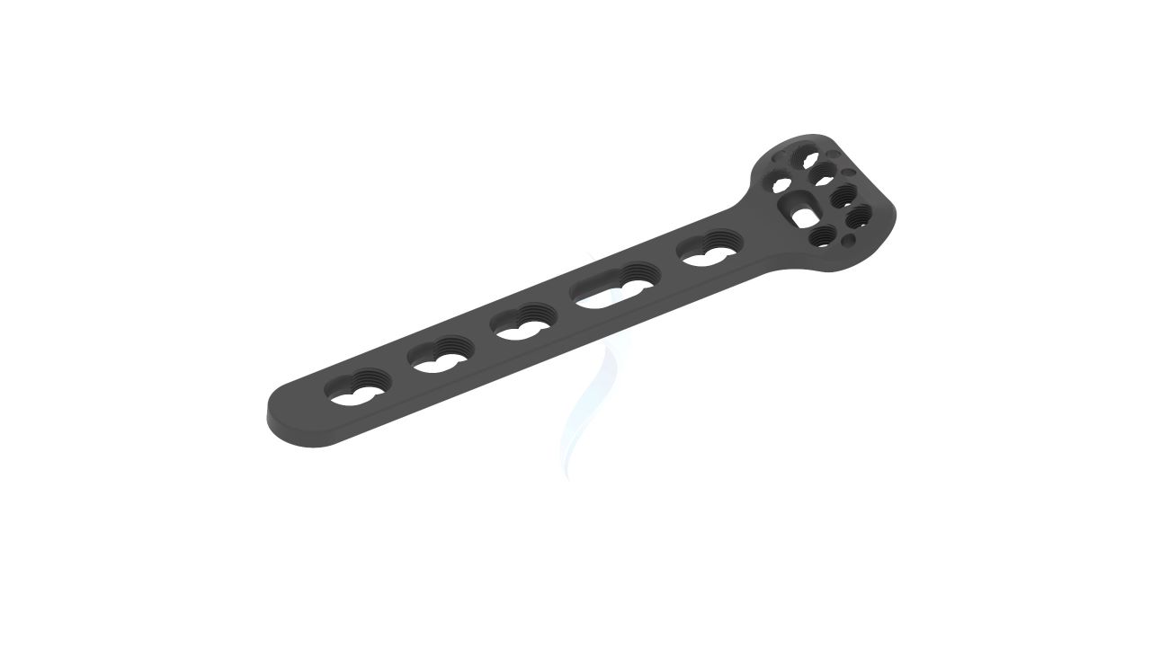
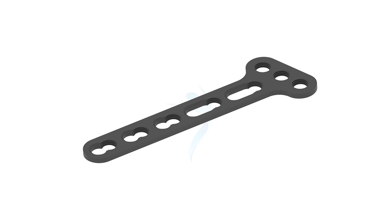
Volar Distal Radius Plate - T Plate (Right Angled)
Product Overview
Introducing our revolutionary Volar Distal Radius T Plate (Right Angled) , a game-changer in orthopedic surgery. Designed to enhance precision and stability during fracture fixation and osteotomy stabilization procedures, this cutting-edge device sets new standards in orthopedic care. Crafted with meticulous attention to detail, the Volar Distal Radius T Plate (Right Angled) is tailored for the volar (palm-side) aspect of the distal radius bone, ensuring optimal alignment and supporting accelerated healing. Its innovative design, coupled with high-quality materials, guarantees durability and reliability, empowering surgeons to deliver exceptional patient outcomes.
Product Uses
- Distal Radius Fracture Fixation : The Volar Distal Radius T Plate (Right Angled) is specifically designed for precise fixation of distal radius fractures, providing stability to promote proper healing.
- Osteotomy Stabilization :Surgeons can utilize the plate to stabilize the bone after an osteotomy, ensuring accurate alignment and support during the healing process.
- Malunion Correction : The plate can be employed to correct malunions (improperly healed fractures) of the distal radius, restoring proper alignment and function.
- Joint Arthrodesis : For cases requiring joint fusion, the Volar Distal Radius T Plate (Right Angled) can be used to immobilize the joint and facilitate the fusion process.
- Corrective Surgery for Deformities : The plate is valuable in corrective surgeries for deformities of the distal radius, aiding in realigning the bone to its anatomically correct position.
Product Specification
- Material :titanium alloy for excellent biocompatibility and strength.
- Size Options : Available in various sizes to accommodate different patient anatomies and surgical requirements.
- Angled Design : Specifically designed with a right-angled configuration to optimize fixation in the volar (palm-side) aspect of the distal radius bone.
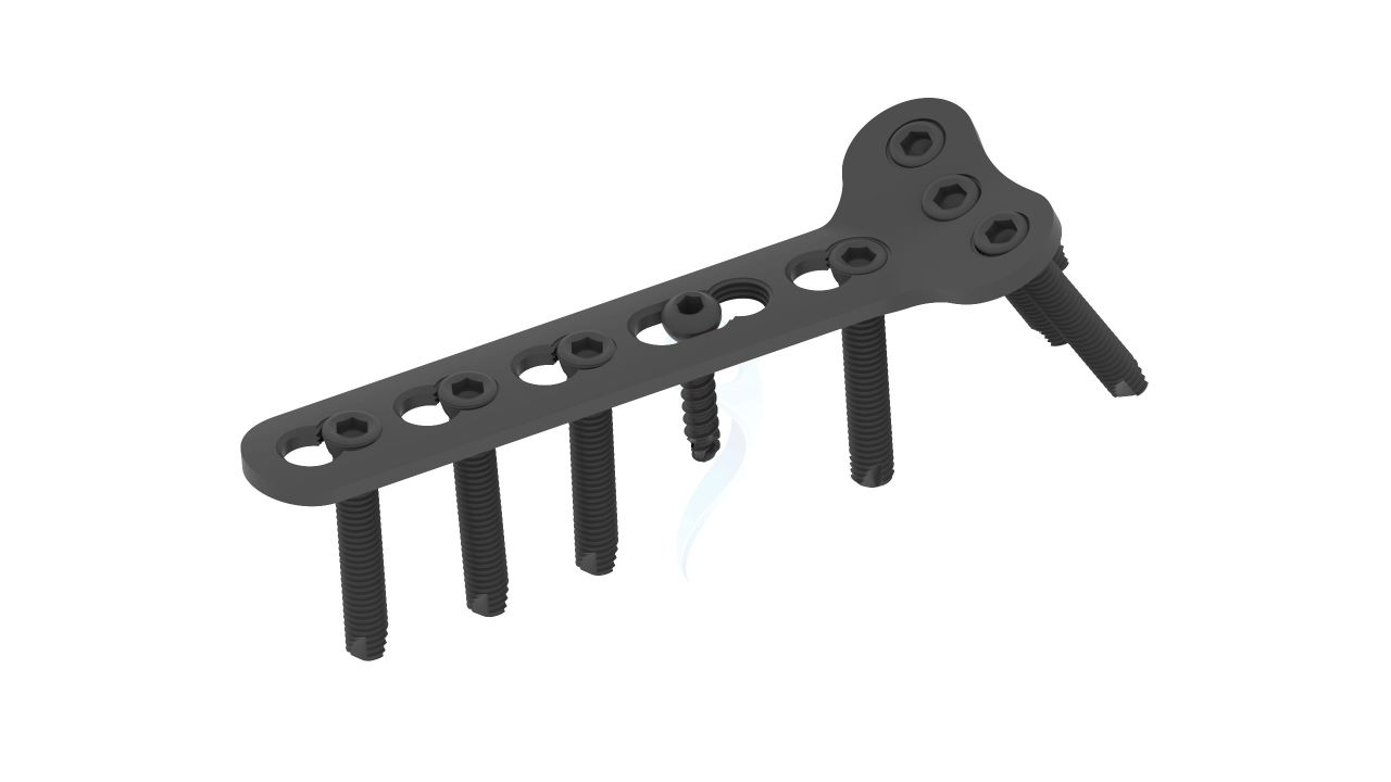
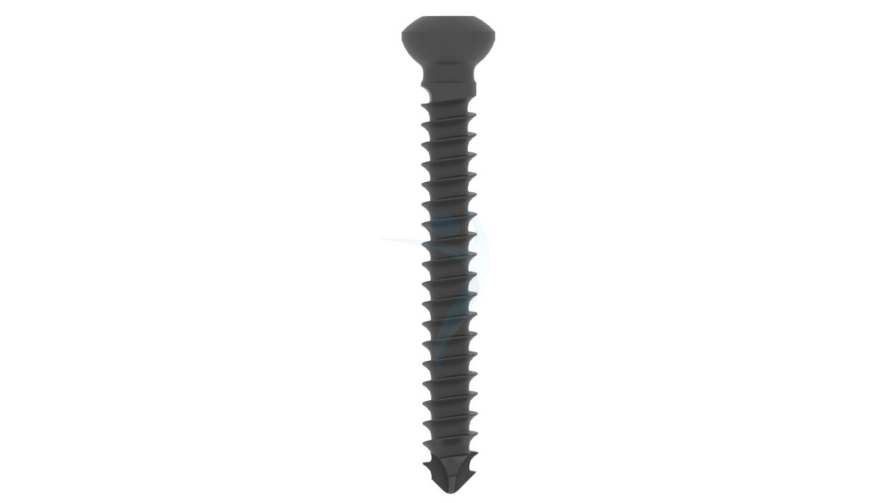
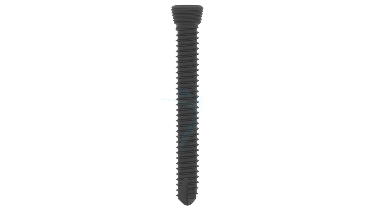
- Screw Holes : Multiple screw holes of varying diameters and lengths to allow for versatile screw placement and stable fixation.
- Locking Mechanism : Featuring a locking system to enhance stability and prevent screw backout, ensuring long-term implant integrity.
- Low Profile : Streamlined and low-profile design to minimize soft tissue irritation and improve patient comfort during the healing process.
Volar Distal Radius - T Plate (Right Angled) Sizes


| Product Code | Name | Material | Head Hole | Shaft Hole |
|---|---|---|---|---|
| PT236.33 | 3.5 T-plate , right-angled , shaft 3 holes, head 3 holes - ASLP | Titanium | 03 | 03 |
| PT236.35 | 3.5 T-plate , right-angled , shaft 5 holes, head 3 holes - ASLP | Titanium | 03 | 05 |
| PT236.44 | 3.5 T-plate , right-angled , shaft 4 holes, head 4 holes - ASLP | Titanium | 04 | 04 |
| PT236.46 | 3.5 T-plate , right-angled , shaft 6 holes, head 4 holes - ASLP | Titanium | 04 | 06 |
Comprehensive Guide for Volar Distal Radius - T Plate (Right Angled)
- Patient Evaluation : Conduct a thorough assessment of the patient's medical history, including any previous injuries or surgeries, allergies, and existing medical conditions.
- Imaging Studies : Review and analyze X-rays, CT scans, or MRI images of the patient's distal radius to determine the extent of the fracture or osteotomy and plan for appropriate plate size and placement.
- Surgical Planning : Collaborate with the surgical team to create a comprehensive surgical plan, considering the fracture type, bone quality, and the desired alignment of the distal radius.
- Implant Selection : Select the appropriate size and configuration of the "Volar Distal Radius T Plate (Right Angled)" based on the patient's anatomy and the surgical plan.
- Informed Consent : Obtain informed consent from the patient, explaining the surgical procedure, potential risks, benefits, and possible alternatives to using the "Volar Distal Radius T Plate (Right Angled)."
- Patient Preparation : Administer the appropriate anesthesia to the patient, ensuring they are comfortable and ready for the surgery. Position the patient correctly, and drape the surgical site in a sterile manner.
- Incision and Exposure : Make an appropriate incision over the volar aspect of the distal radius to expose the fractured or osteotomized bone.
- Fracture Reduction or Osteotomy : If necessary, perform fracture reduction or osteotomy to realign the bone segments and restore the proper anatomy.
- Plate Selection : Select the appropriate size of the "Volar Distal Radius T Plate (Right Angled)" based on preoperative measurements and the patient's anatomy.
- Plate Placement : Carefully position the "Volar Distal Radius T Plate (Right Angled)" on the volar aspect of the distal radius bone, ensuring that the plate's angles match the bone contours accurately.
- Screw Hole Preparation : Use specialized instruments to create pilot holes in the bone through the plate's pre-drilled screw holes. Ensure proper screw trajectory and depth.
- Immediate Postoperative Period : Transfer the patient to the recovery area and closely monitor their vital signs and overall condition.Ensure the patient's comfort and safety during the initial recovery phase.
- Wound Care : Keep the surgical incision site clean and dry.Change dressings regularly, following the surgeon's instructions or institutional protocols.
- Immobilization and Weight-Bearing : Instruct the patient to follow weight-bearing guidelines, if applicable, as recommended by the surgeon.Provide guidelines for immobilization, which may include the use of a cast, splint, or brace to support the wrist during the initial healing phase.
- Physical Therapy and Rehabilitation : Initiate a guided physical therapy program as prescribed by the surgeon to promote wrist mobility and strength gradually.
- Activity Restrictions : Instruct the patient to avoid strenuous activities and heavy lifting during the early postoperative period to protect the healing bone and implant.

.png "Femoral Neck System (FNS)")


