Volar Distal Radius Plate - 2.4/2.7 mm Variable Angle Rim
Product Overview
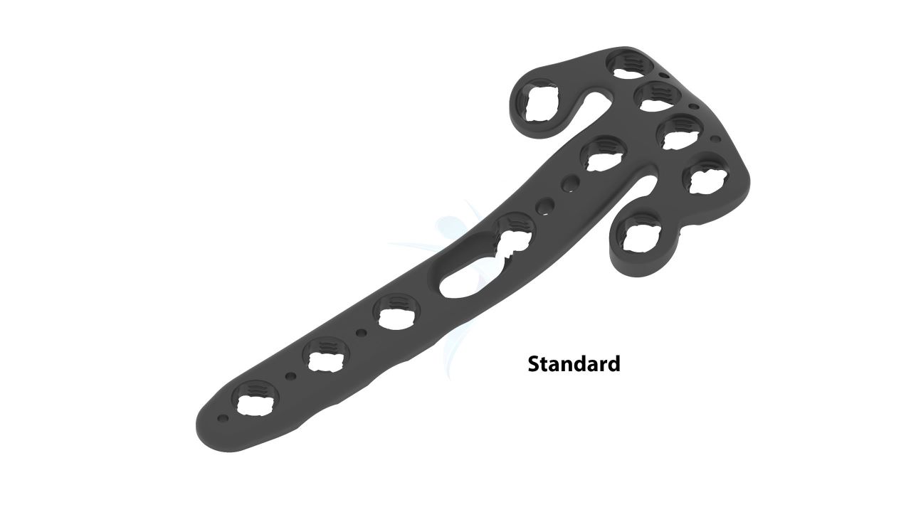
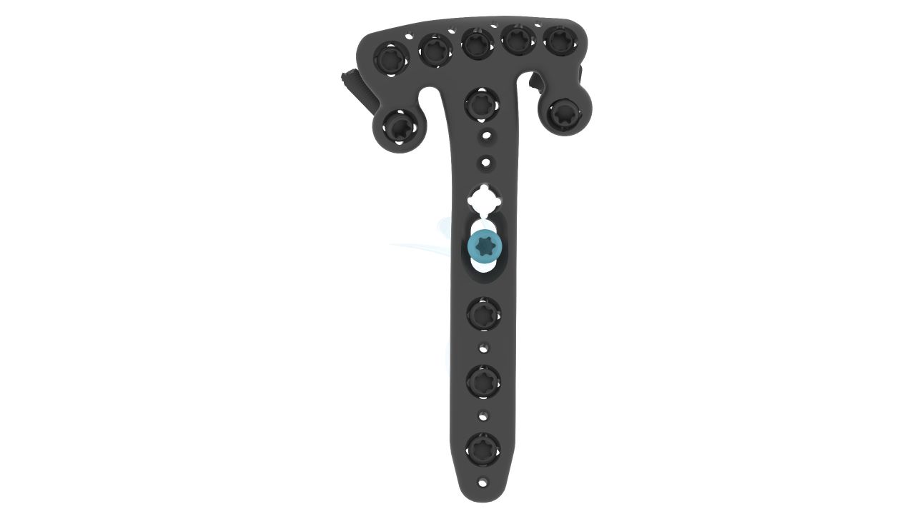
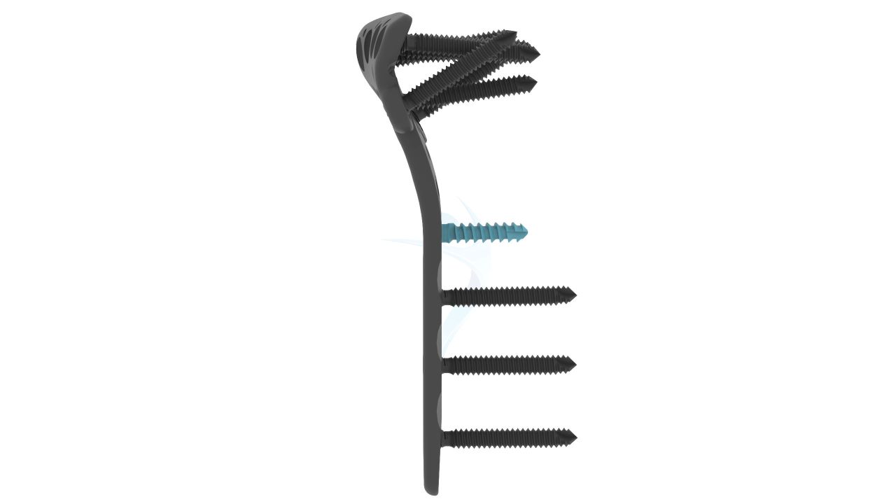
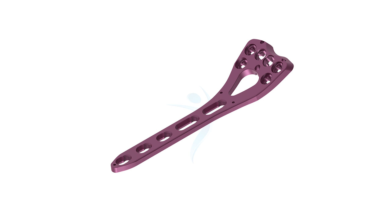
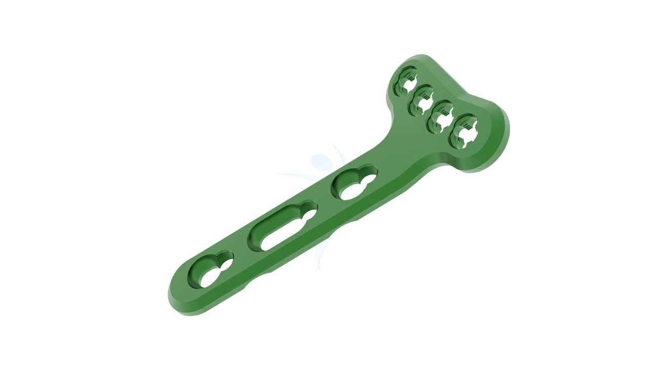
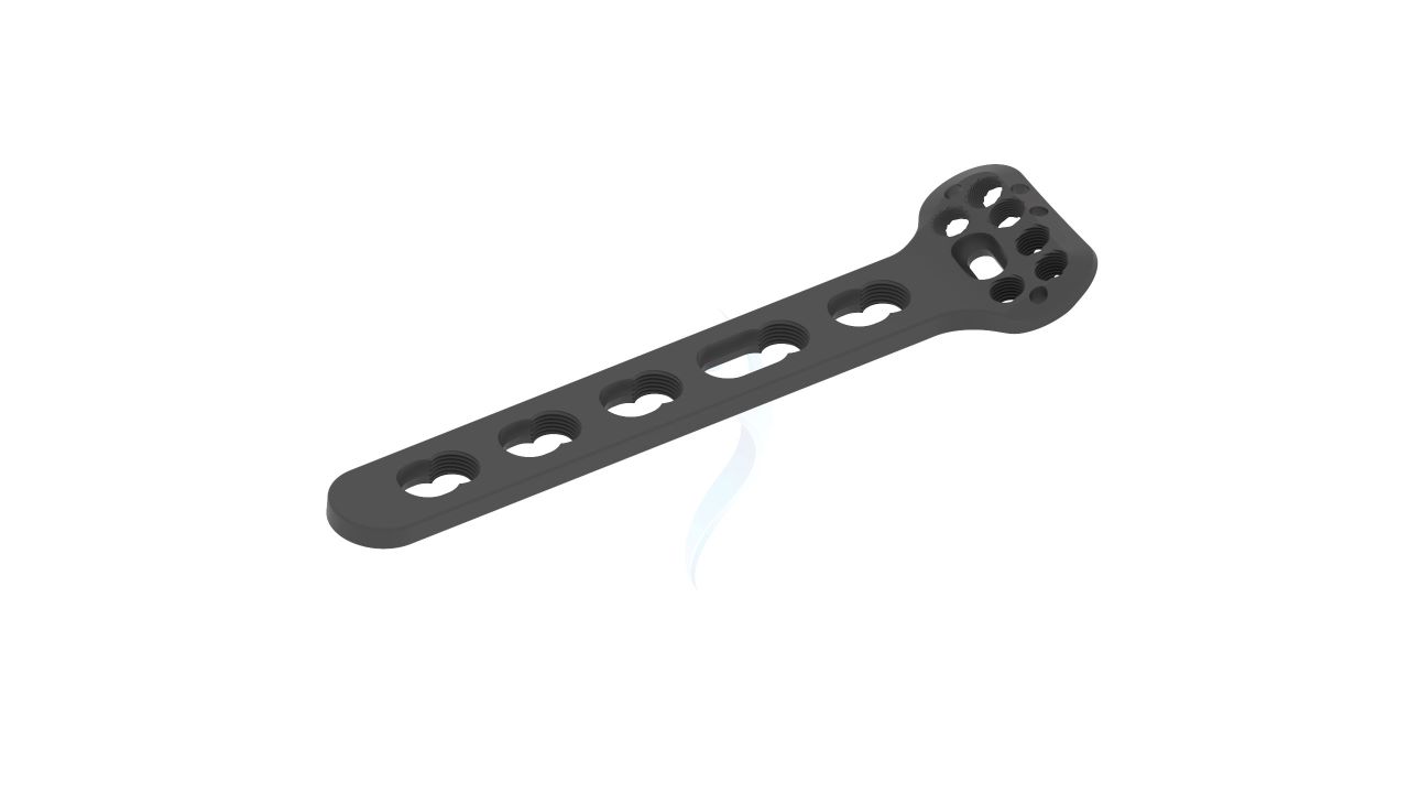
Introducing our advanced Volar Distal Radius Rim Plate (Variable Angle 2.4/2.7 mm) , a cutting-edge orthopedic implant designed to redefine wrist fracture repair. This innovative plate offers adjustable angle fixation, empowering surgeons to provide tailored solutions for even the most complex cases. Precision-crafted and engineered for stability, our plate sets a new standard in orthopedic care. Elevate patient outcomes and ensure the utmost in fracture healing and wrist function restoration with this trusted implant.

Product Uses
- Distal Radius Fracture Fixation :The primary purpose is to stabilize and fixate distal radius fractures, providing support and alignment for optimal healing.
- Variable Angle Fixation : The plate's adjustable angle feature allows surgeons to adapt the fixation angle to the specific needs of each patient and fracture, enhancing surgical flexibility.
- Complex Fractures : It is particularly valuable in cases involving complex or comminuted distal radius fractures, where stability and precise alignment are critical.
- Fracture Reduction : The plate facilitates the reduction of displaced fractures, assisting in aligning bone fragments accurately.
- Wrist Joint Stabilization : By providing stability to the wrist joint, it helps prevent post-operative deformities and supports functional recovery.
Product Specification
- Size : The plate has a width of 2.4mm, ensuring stability while accommodating varying anatomical structures.
- Material :Manufactured from high-quality medical-grade titanium alloy for biocompatibility, strength, and durability.
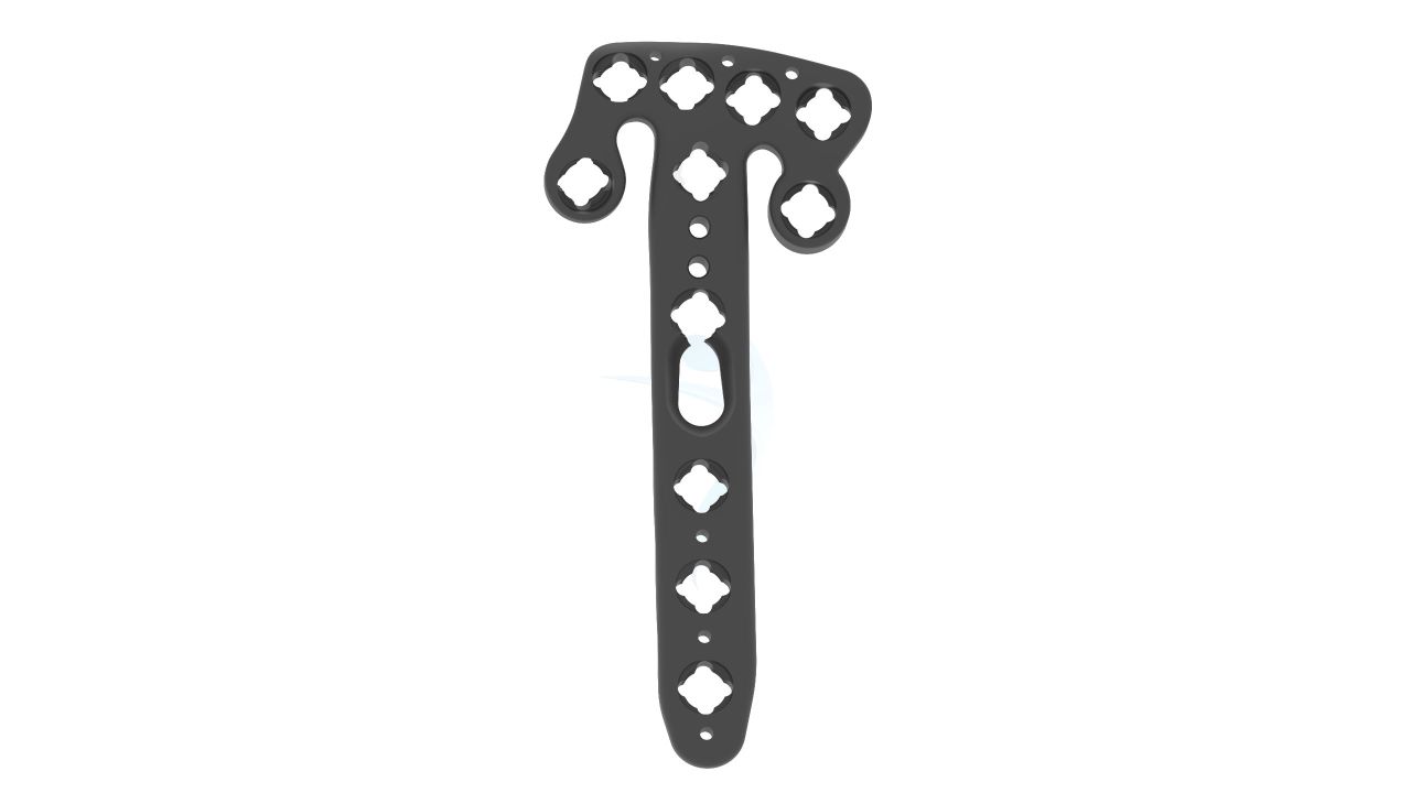
- Design : The plate features a variable angle design, allowing surgeons to adjust the angle of fixation during surgery to suit the patient's specific needs.
- Thickness : The plate's thickness is standardized at 2.4mm to maintain structural integrity.
- Thickness : The plate's thickness will correspond to the selected width, with one side being 2.4mm thick and the other side 2.7mm thick, ensuring strength and stability.
Volar Distal Radius - Rim Plate (Variable Angle 2.4/2.7 mm) Sizes


| Product Code | Name | Material | Head Hole | Shaft Hole | Angled |
|---|---|---|---|---|---|
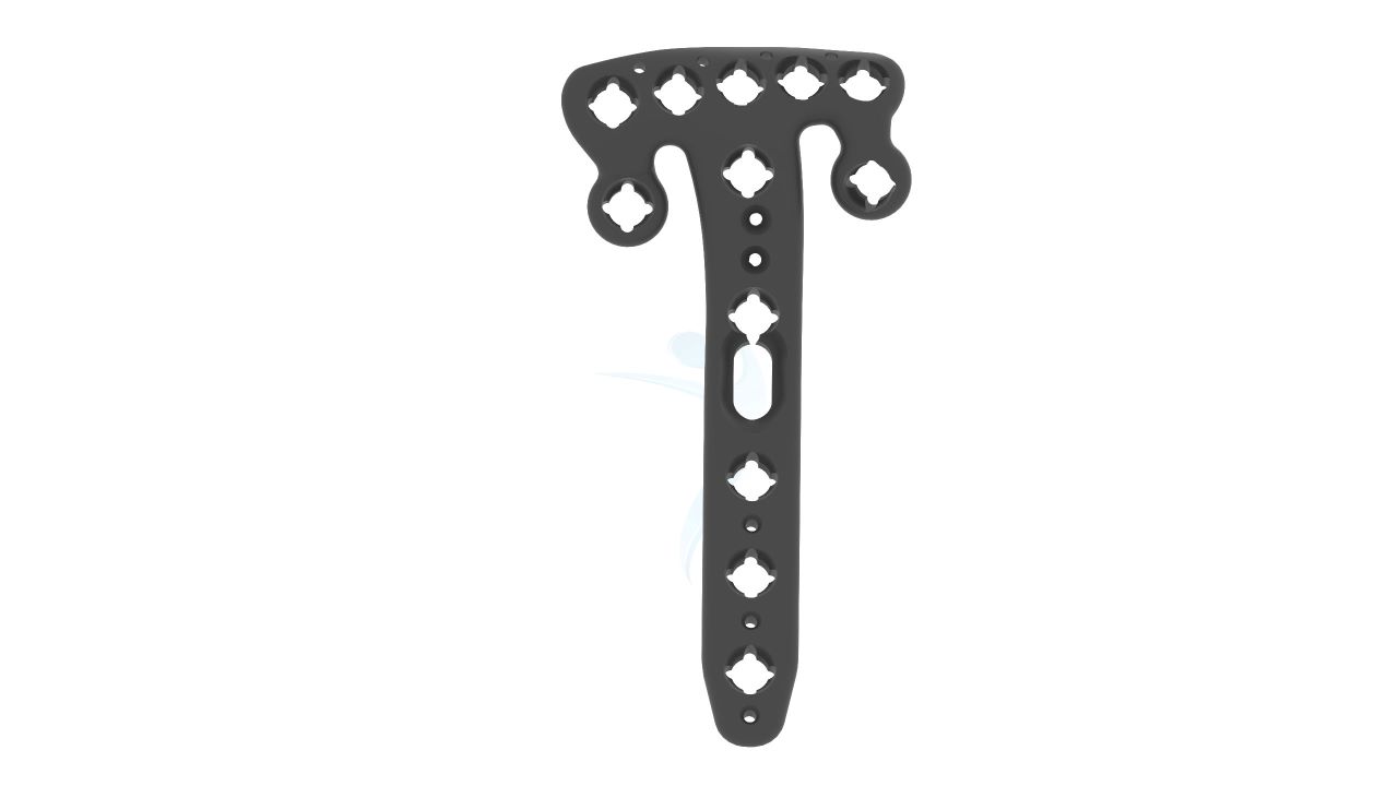
| PT319.05.01 | 2.4/2.7 Distal radius volar Rim Plate Standard 5 hole Right - VALP | Titanium | 7 | 5 | Right |
| PT319.05.02 | 2.4/2.7 Distal radius volar Rim Plate Standard 5 hole Left - VALP | Titanium | 7 | 5 | Left |
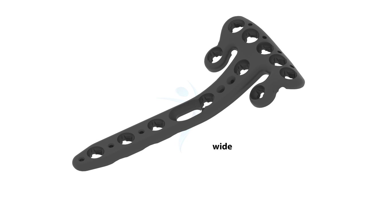
| PT320.05.01 | 2.4/2.7 Distal Radius Volar Rim Plate Wide 5 Hole Right - VALP | Titanium | 7 | 5 | Right |
| PT320.05.02 | 2.4/2.7 Distal Radius Volar Rim Plate Wide 5 Hole Left - VALP | Titanium | 7 | 5 | Left |
Comprehensive Guide for Volar Distal Radius Plate - 2.4/2.7 mm Variable Angle Rim
- Patient Evaluation : The surgeon conducts a comprehensive evaluation of the patient's medical history, wrist condition, and any previous surgeries.
- Imaging : X-rays, CT scans, or other imaging studies are performed to assess the extent and nature of the distal radius fracture.
- Surgical Planning : Based on imaging results, the surgeon plans the procedure, including the selection of the appropriate plate size (2.4mm), length, and the anticipated angle of fixation using the variable angle feature.
- Informed Consent : The surgeon discusses the procedure, potential risks, and benefits with the patient, obtaining informed consent.
- Anesthesia : Anesthesia options (local, regional, or general) are discussed with the patient and administered as per the patient's and surgeon's preferences.
- Patient Positioning : The patient is positioned on the operating table with the affected wrist accessible and exposed.
- Incision : A sterile field is established, and a carefully planned incision is made over the distal radius. The incision's length and location are determined by the fracture pattern and the chosen plate size.
- Fracture Reduction : The surgeon carefully manipulates and reduces the fractured bone fragments, ensuring proper alignment.
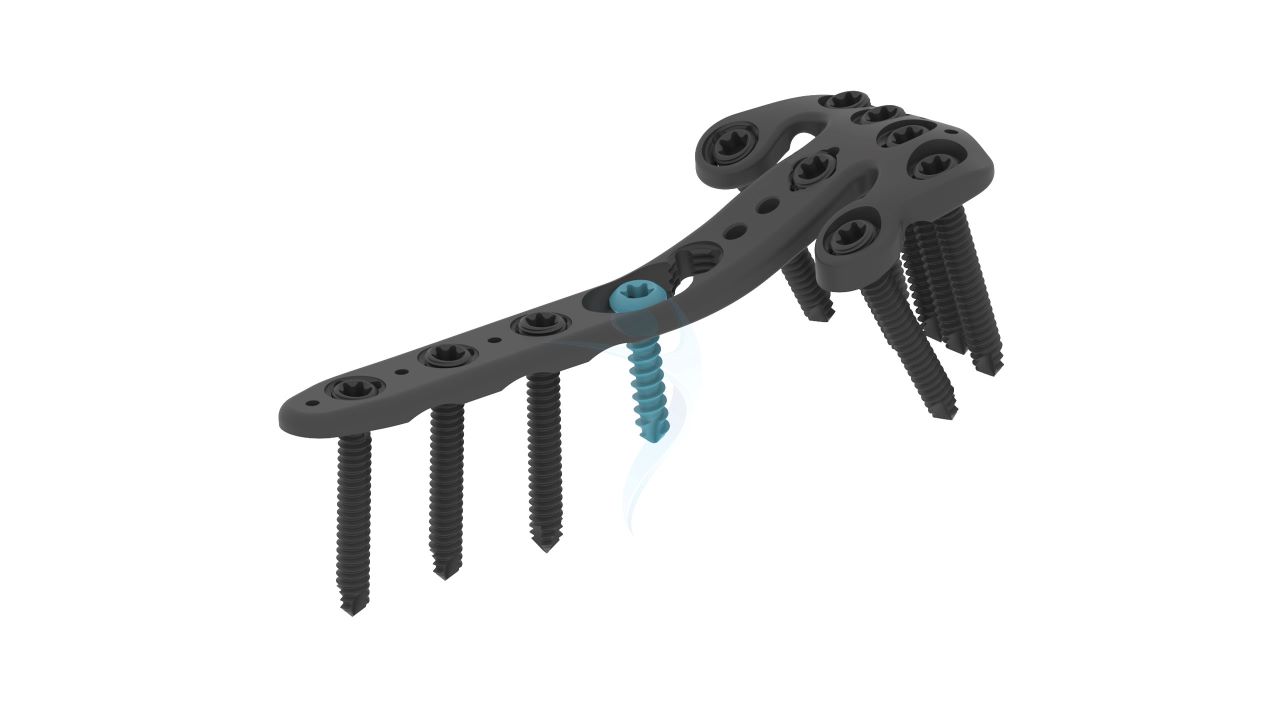
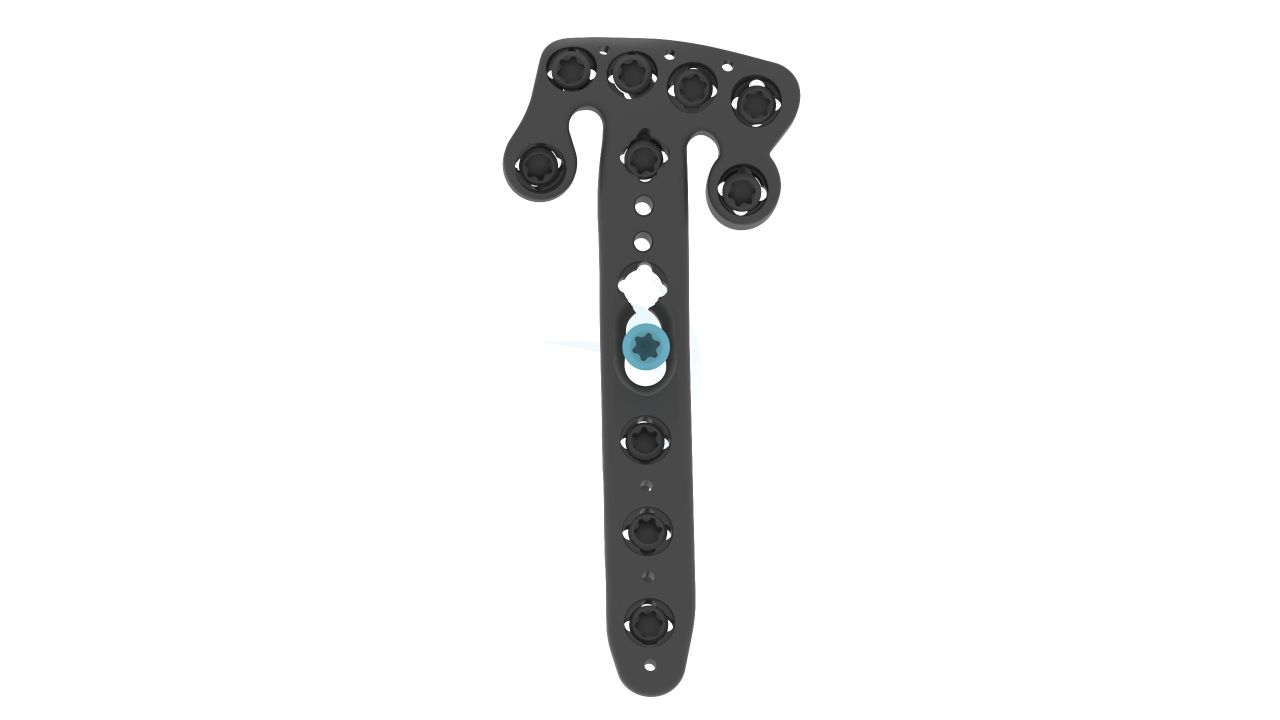
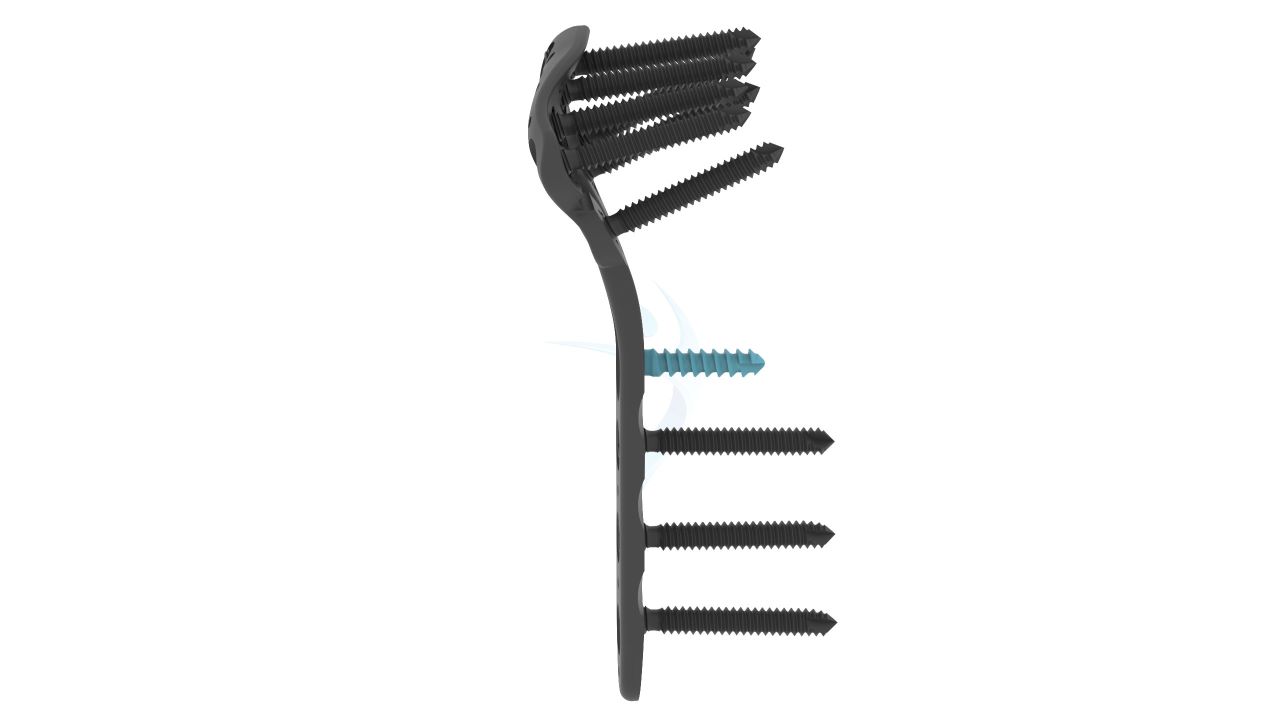
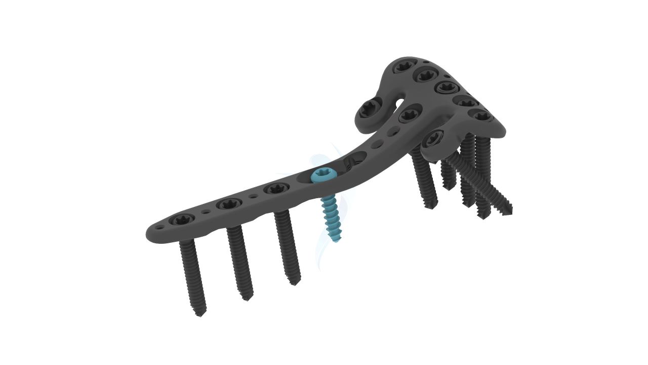
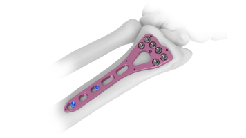
- Plate Placement : The "Volar Distal Radius Rim Plate (Variable Angle 2.4/2.7 mm)" is positioned over the fracture site, and screws are inserted through the plate's screw holes to secure it in place. The variable angle feature allows for fine-tuned placement.
- Variable Angle Fixation : Utilizing the variable angle feature of the plate, the surgeon adjusts the angle of fixation as needed to achieve optimal alignment and stability.
- Wound Closure : The incision is meticulously closed using sutures or staples, ensuring a clean and infection-free wound closure.
- Recovery Room : The patient is monitored in the recovery room to ensure stable vital signs and absence of immediate post-operative complications.
- Pain Management : Pain management protocols are initiated as needed to keep the patient comfortable.
- Physical Therapy : Depending on the surgeon's recommendations, physical therapy and rehabilitation exercises may be initiated to promote wrist mobility and strength.
- Follow-Up Appointments : The patient is scheduled for follow-up appointments with the surgeon to monitor the healing process, remove sutures or staples, and assess overall recovery.
- X-ray Monitoring : Regular X-rays are taken to evaluate bone healing and the position of the implant.

.png "Femoral Neck System (FNS)")


