Elbow Plate (ASLP)- 3.5 Olecranon
Product Overview
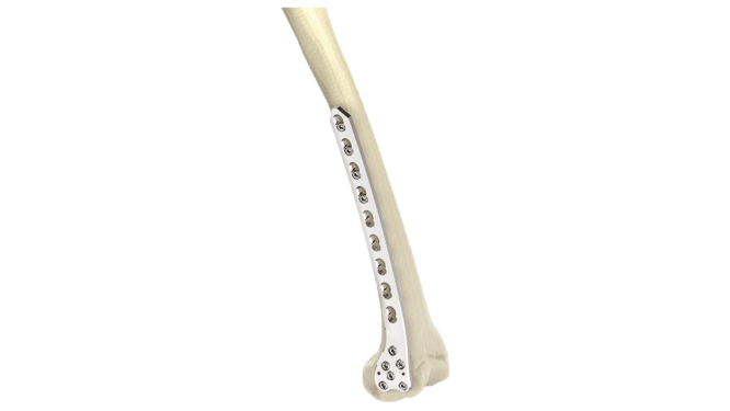
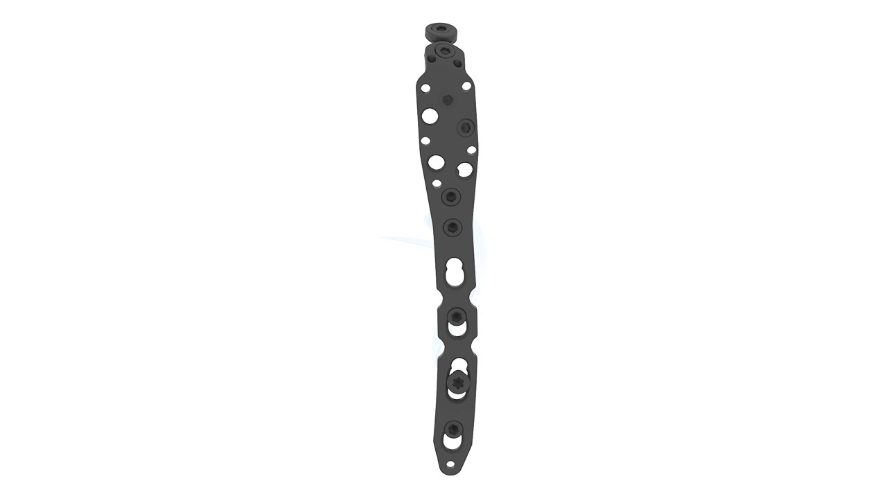
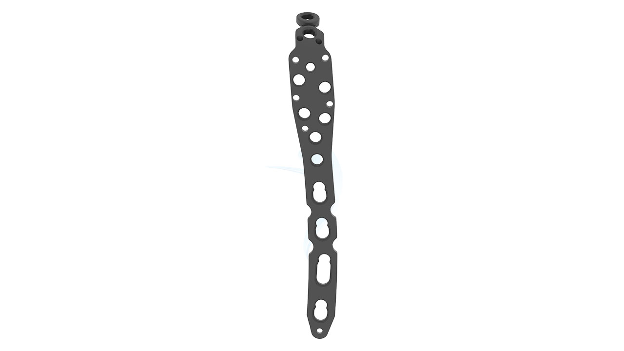
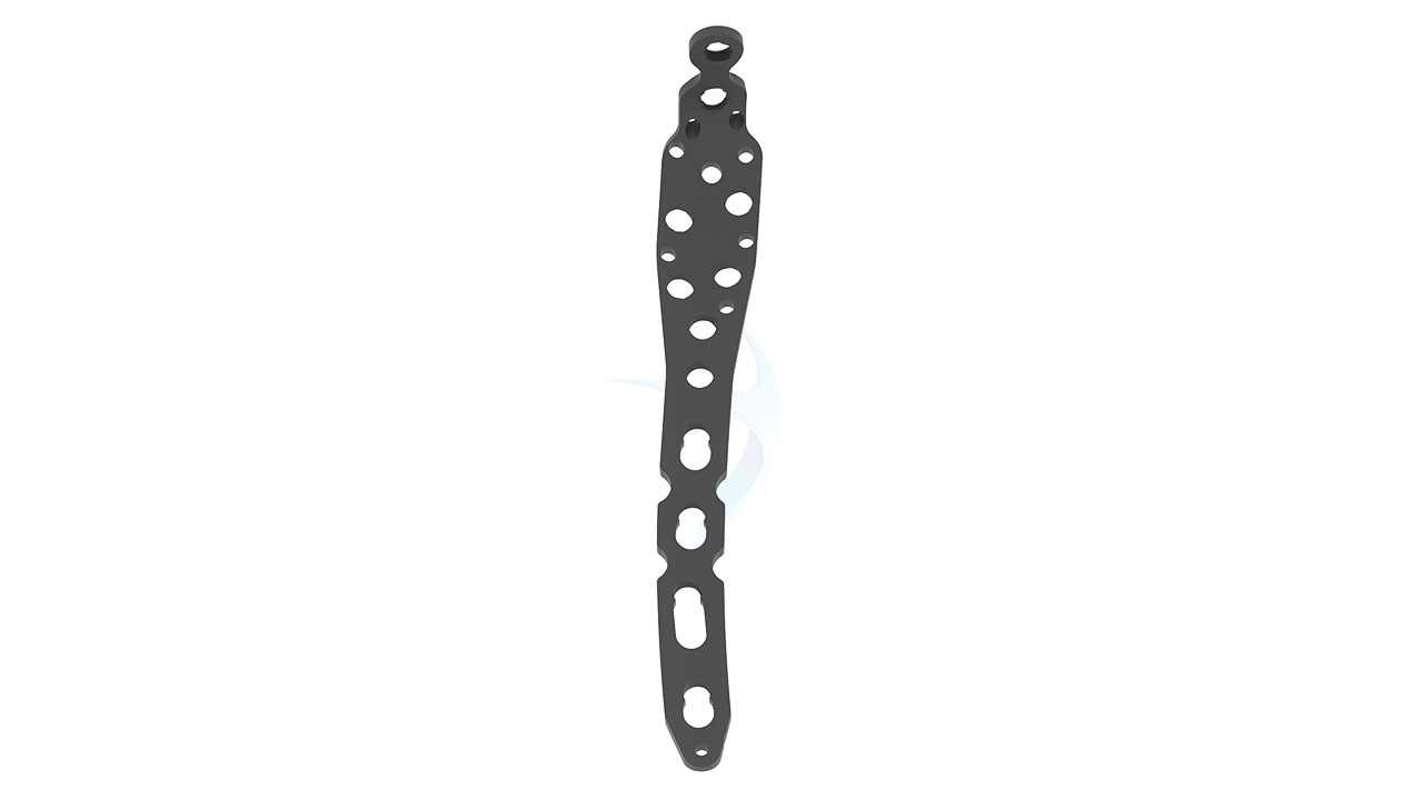
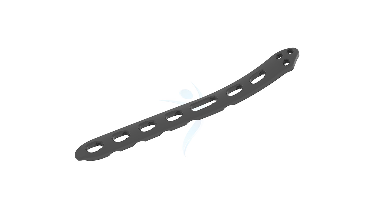
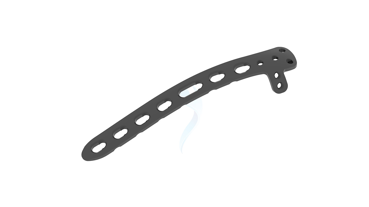
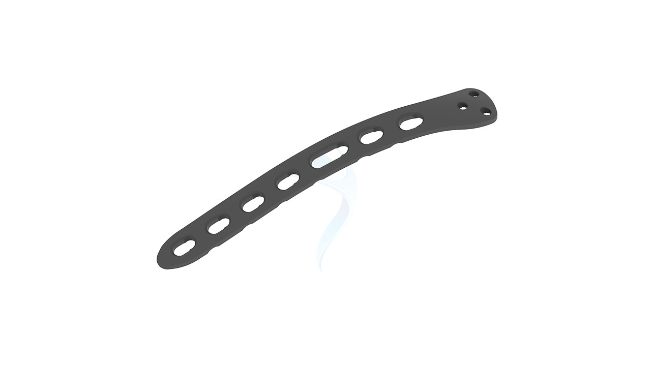
The Elbow Plate ASLP - 3.5 Olecranon is an advanced orthopedic implant designed to provide stability and support for fractures and injuries affecting the olecranon, the prominent bony prominence at the tip of the elbow. With its specialized design and precision engineering, this plate offers secure fixation and promotes optimal healing, restoring function to the elbow joint. Whether used in trauma cases or elective procedures, the 3.5 Olecranon Plate represents a reliable solution for orthopedic surgeons seeking to address elbow pathologies with confidence and precision.
Product Uses
- Olecranon Fractures : This implant is primarily used to treat fractures of the olecranon, the bony prominence at the tip of the elbow. It provides stability and support to the fractured bone fragments, allowing for proper alignment and healing.
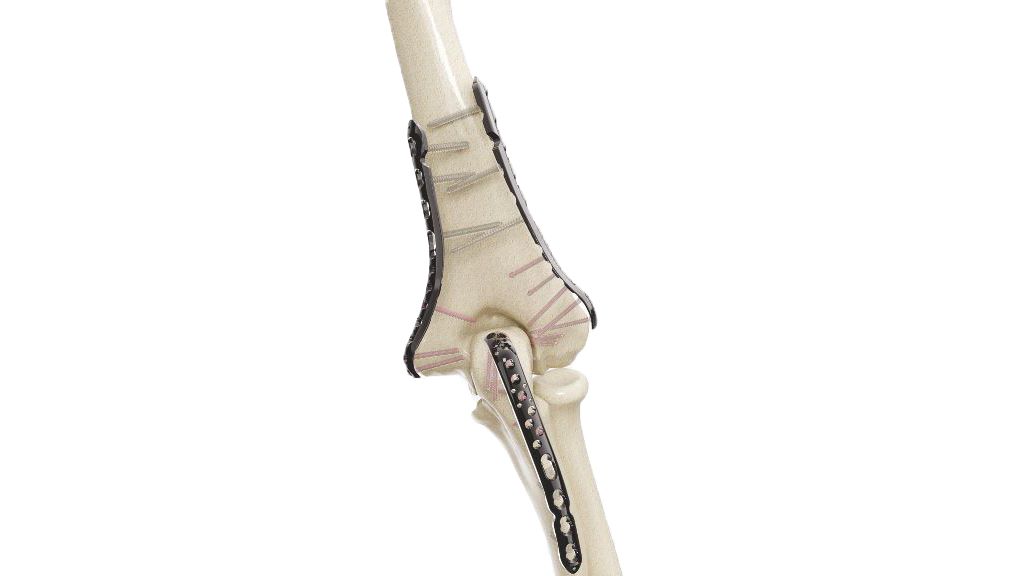
- Fractures Involving the Proximal Ulna : In addition to olecranon fractures, the Elbow Plate ASLP - 3.5 Olecranon can be used to stabilize fractures involving the proximal portion of the ulna bone, which forms the elbow joint.
- Osteotomies and Corrective Surgeries : Orthopedic procedures involving osteotomies or corrective surgeries around the elbow joint may utilize this implant to maintain the corrected position and promote proper healing.
- Nonunions and Malunions : In cases where fractures fail to heal (nonunion) or heal in an incorrect position (malunion), revision surgery may be necessary, and this implant can aid in realigning and stabilizing the bone fragments.
- Trauma : The Elbow Plate ASLP - 3.5 Olecranon can be used to stabilize traumatic injuries to the elbow joint, such as fractures resulting from falls, sports injuries, or motor vehicle accidents.
Product Specification
- Material : Typically made of biocompatible materials such as titanium or stainless steel, chosen for their strength, durability, and compatibility with the human body.
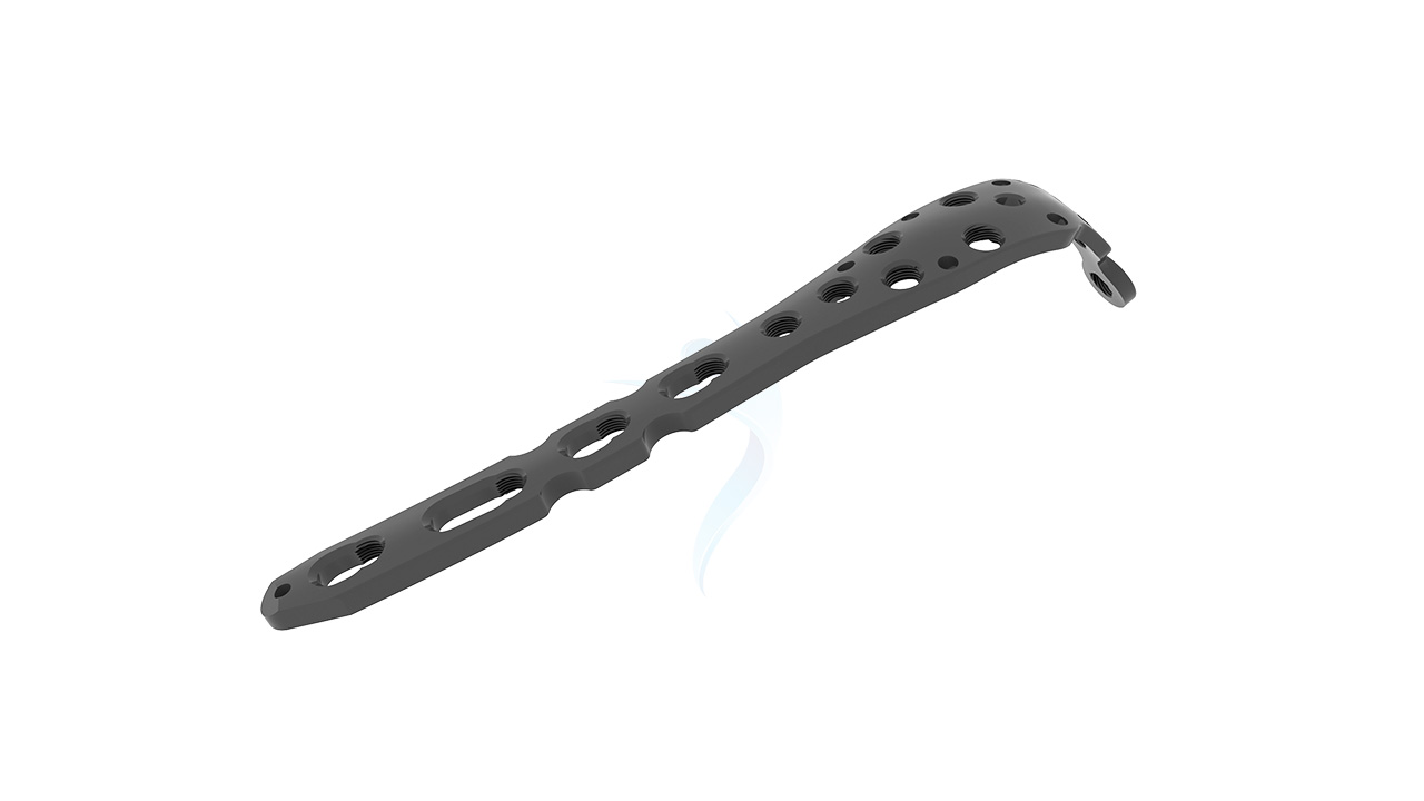
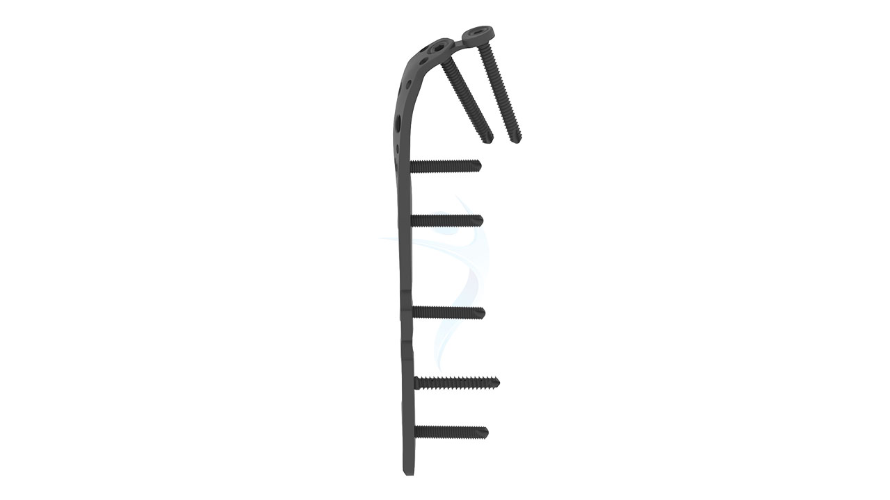
- Plate Design : Specifically designed to fit the anatomy of the olecranon, the bony prominence at the tip of the elbow.
- Plate Thickness : Available in a thickness of 3.5 mm, chosen to provide adequate strength and stability for fractures and injuries affecting the olecranon.
- Plate Length : The length of the plate may vary depending on the size of the patient's anatomy and the specific requirements of the surgical procedure. Typically designed to cover the length of the olecranon and provide sufficient surface area for screw fixation.
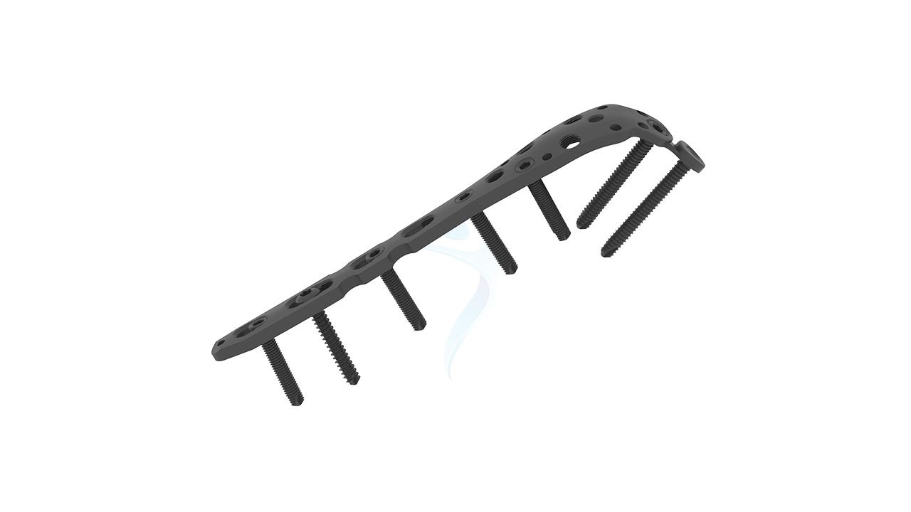
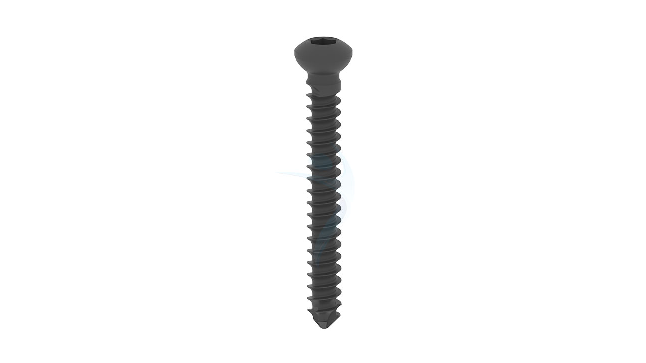
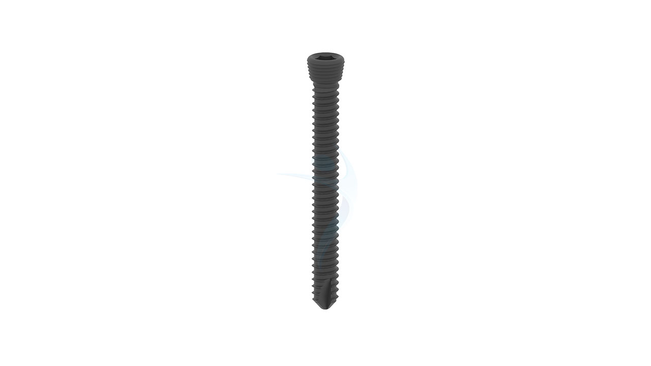
- Screw Holes : Features multiple screw holes along its length, allowing for flexible fixation with orthopedic screws. Strategically positioned to facilitate optimal bone purchase and secure fixation, even in complex fracture patterns.
Elbow Plate (ASLP) - 3.5 Olecranon Sizes
Comprehensive Guide for Elbow Plate (ASLP) - 3.5 Olecranon
- Patient Evaluation : The patient's medical history, including any previous surgeries or medical conditions, is reviewed. Imaging studies such as X-rays, CT scans, or MRI scans are performed to assess the extent of the elbow injury or condition and aid in surgical planning.
- Surgical Planning : The surgeon evaluates the fracture or condition of the olecranon and surrounding structures. The appropriate size and thickness of the implant are determined based on the patient's anatomy and the specific requirements of the surgical procedure.
- Patient Preparation : The patient receives instructions on pre-operative preparations, which may include fasting prior to surgery, discontinuation of certain medications, and pre-operative skin preparation.
- Consent and Education : The surgeon discusses the planned procedure, potential risks, expected outcomes, and alternative treatment options with the patient. The patient has the opportunity to ask questions and provide informed consent for the surgery.
- Anesthesia : The patient is placed under general anesthesia to ensure comfort and immobility during the surgical procedure. Regional anesthesia techniques may also be used to provide additional pain control during and after surgery.
- Incision : The surgeon makes an incision over the elbow joint, typically along the posterior aspect, to access the olecranon and the fracture site.
- Fracture Reduction : If the olecranon fracture fragments are displaced, the surgeon carefully manipulates them into proper alignment (reduction) to restore normal anatomy and alignment of the elbow joint.
- Plate Placement : The Elbow Plate ASLP - 3.5 Olecranon is carefully positioned over the fracture site, with its pre-contoured shape matching the natural curvature of the olecranon. The plate is secured to the bone using orthopedic screws, which are inserted through the plate's screw holes into the bone to provide stable fixation.
- Closure : Once the plates are securely in place and the fracture is stabilized, the incision is closed using sutures or staples. Sterile dressings are applied to the surgical site to promote healing and reduce the risk of infection.
- Recovery Room : The patient is monitored closely in the recovery room as they wake up from anesthesia. Pain management medications are administered as needed to ensure comfort during the initial
- Physical Therapy : Depending on the surgeon's recommendations and the patient's condition, physical therapy may begin soon after surgery to promote mobility, strength, and range of motion in the elbow joint. The therapist will guide the patient through exercises designed to support healing and prevent stiffness or muscle weakness.
- Follow-Up Care : The patient will have regular follow-up appointments with the surgeon to monitor healing progress, assess the function of the implant, and remove sutures or staples as needed. X-rays may be taken at these appointments to evaluate bone healing and implant stability.
- Activity Restrictions : The patient will be instructed on activity restrictions and proper care of the surgical site to minimize the risk of complications and support optimal healing. Gradual rehabilitation exercises will be prescribed to gradually increase strength and mobility in the elbow joint while avoiding excessive stress on the implant or fracture site.
- Long-Term Management : Long-term management may involve ongoing monitoring of the implant, periodic imaging studies, and adjustments to the rehabilitation program as needed.




.png)

